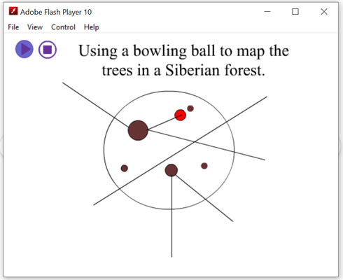
(A) Bowling balls passing through a tree forest. Tree trunks of different sizes are depicted by brown circles.
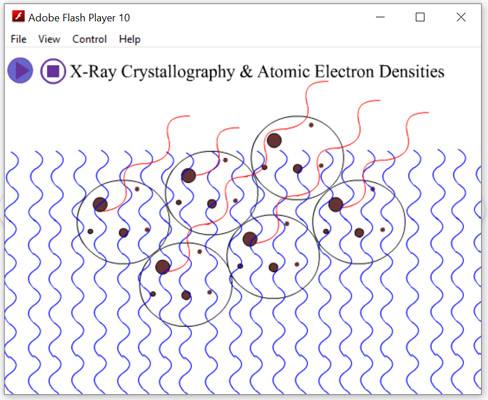
(B) X-ray diffraction by a homogenous crystallized lattice of molecules.
Different atomic nuclei are depicted by brown circles.
(E) Single particle cryo-EM image processing workflow. Extended Data Fig. 5 . Z. Ke et al. Nature (2020) 588 pp. 498–502.
(F) Structural analysis of SARS-CoV-2 spike trimers on intact virions.
Fig. 2. Z. Ke et al. Nature 588 pp. 498–502 (2020).
Fig. 2. Z. Ke et al. Nature 588 pp. 498–502 (2020).


(C) X‐ray photograph of a sperm whale myoglobin crystal.
J. Kendrew and M. Perutz, (late 1950s).
J. Kendrew and M. Perutz, (late 1950s).
(D) Myoglobin alpha-helical electron density map.
Kendrew and Perutz (late 1950s).
Kendrew and Perutz (late 1950s).

